|
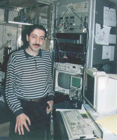
 Maryland setup |
|
|
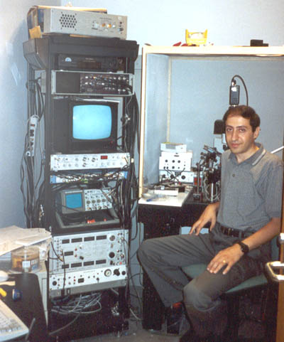
Maryland setup |
 The specific pattern of electrical activity observed in a neuron is dictated
by the complement of ion channels expressed by the cell and by the synaptic
inputs that it receives. Chemicals in the brain (that is, neurotransmitters
and drugs) change those patterns of activity, often by modulating the behavior
of ion channels postynaptically or by presynaptic modulation of neurotransmitter
release. My research focuses on investigating catecholaminergic modulation
of neuronal excitability through direct actions on membrane properties
or by modulating excitatory and inhibitory synaptic release. I have been
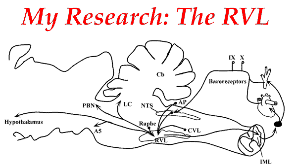
interested in studying the function of the noradrenergic system at the
cellular level in two brain structures: the rostral ventrolateral medulla
and the olfactory bulb.
The specific pattern of electrical activity observed in a neuron is dictated
by the complement of ion channels expressed by the cell and by the synaptic
inputs that it receives. Chemicals in the brain (that is, neurotransmitters
and drugs) change those patterns of activity, often by modulating the behavior
of ion channels postynaptically or by presynaptic modulation of neurotransmitter
release. My research focuses on investigating catecholaminergic modulation
of neuronal excitability through direct actions on membrane properties
or by modulating excitatory and inhibitory synaptic release. I have been
interested in studying the function of the noradrenergic system at the
cellular level in two brain structures: the rostral ventrolateral medulla
and the olfactory bulb.
 Moreover, neurons in the C1 area initiate the cardiovascular response to
ischemia, and mediate rapid sympathoexcitatory pressor response to acute
hypoxia. On the other hand, there is now good evidence that the key mechanisms
for breathing rhythm generation resides within the RVL where several neuron
types likely to support the rhythm generating process have been identified.
Moreover, neurons in the C1 area initiate the cardiovascular response to
ischemia, and mediate rapid sympathoexcitatory pressor response to acute
hypoxia. On the other hand, there is now good evidence that the key mechanisms
for breathing rhythm generation resides within the RVL where several neuron
types likely to support the rhythm generating process have been identified.
 I have investigated pre- and postsynaptic actions of opioid and adrenergic
receptor agonists (including centrally-acting antihypertensive drugs) on
RVL bulbospinal neurons in brainstem slices. My major findings were the
following:
I have investigated pre- and postsynaptic actions of opioid and adrenergic
receptor agonists (including centrally-acting antihypertensive drugs) on
RVL bulbospinal neurons in brainstem slices. My major findings were the
following:
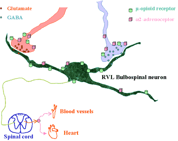
The majority of C1 and non-C1 RVL bulbospinal neurons are inhibited by met-enkephalin via activation of µ-opioid receptors. The inhibition of RVL neurons by opioids occurs: - Postsynaptically: Opening of inwardly rectifying potassium channels. - Presynaptically: Decrease of glutamatergic transmission. These mechanisms could explain the hypotensive and sympathoinhibitory actions of opioids transmitters in the RVL in particular during hypotensive hemorrhage. NE inhibits bulbospinal RVL neurons by activating alpha2-adrenoceptors (alpha2-ARs) located postsynaptically and presynaptically on glutamatergic terminals. Contrary to our expectations, alpha2-ARs were also found on GABAergic inputs to these cells. Unlike the postsynaptic effects, the decrease of synaptic transmission by NE was not sensitive to barium, indicating that different mechanisms of action are involved in the pre- and postsynaptic effects of NE. Finally, all effects of NE were mimicked by the prototypical imidazoline ligand moxonidine which was found to behave like an alpha2-AR agonist. |
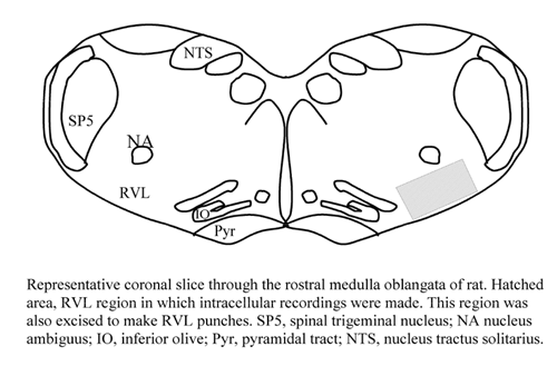
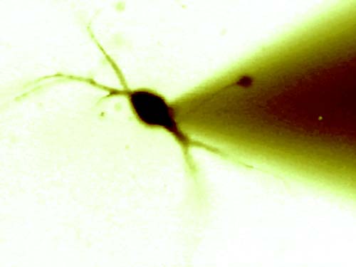 Picture of an RVL neuron recorded using the whole-cell |
![]()
| The main olfactory bulb (MOB) is a highly organized, well characterized, relatively simple cortical network that functions to process olfactory information. It receives a significant modulatory noradrenergic input from the locus coeruleus which densely innervates this network with a higher degree of laminar specificity than in any other forebrain target. Previous in vivo and in vitro work showed that norepinephrine (NE) increases the sensitivity of mitral cells to weak olfactory inputs. The goal of my current research is to investigate the cellular basis for this action of NE in olfactory bulb slices and to identify the type of adrenergic receptor involved in regulating the excitability of mitral cells, the main output of the olfactorybulb. | 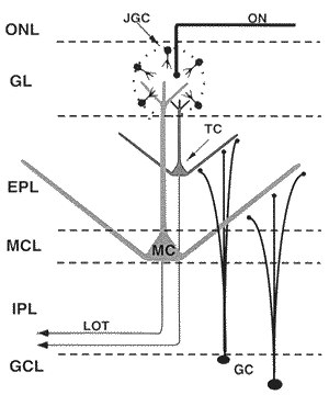 |